Treatment of Back Pain Innovative Methods in Modern Medicine

Treatment of Back Pain: Innovative Methods in Modern Medicine

The treatment of back pain has evolved significantly with the remarkable advances in the field of modern medicine. Medical innovation targets back pain, fostering advanced pain management approaches.

Advancements in diagnosis, interventions, and multidisciplinary care redefine back problem management. Exploring these aspects aids understanding modern medicine’s commitment to personalized back pain solutions

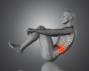
Back pain, pervasive across diverse ages and lifestyles globally, stands as one of the most prevalent medical conditions. Modern medicine has made great strides in understanding and effectively treating back pain, offering patients diverse and innovative ways to manage this debilitating problem.


CONTENT:

  1. The new method for treatment of back pain
  2. The new device for treatment of back pain
  3. Currently recommended treatment for back pain
  4. The most powerful medicine for back pain
  5. The safest pain medication for long-term use

The new method for treatment of back pain

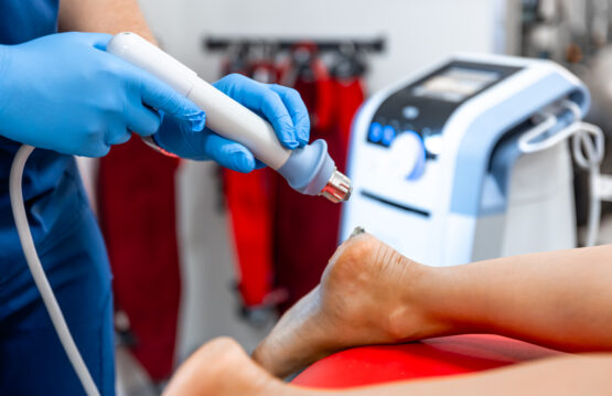
High Intensity Focused Ultrasound (HIFU) therapy represents one of the relatively new methods used to treat back pain. This non-invasive method uses ultrasonic waves to treat back pain-causing conditions such as spinal neuralgia, reducing pain sensations by precisely focusing energy on the affected areas.

By using high frequencies of ultrasound, HIFU can precisely focus energy on specific areas, allowing doctors to treat back pain-causing conditions, such as spinal neuralgia, with minimal invasiveness and without affecting the surrounding tissues. The principle of operation consists in the transmission of ultrasonic waves that turn into heat in specific areas of the spine, helping to reduce inflammation and pain sensations.

This method offers non-invasive treatment, ensuring swift recovery and avoiding complications linked to traditional surgery. Furthermore, HIFU may represent an alternative for those who do not respond to other forms of treatment or who are not ideal candidates for surgery.

Although HIFU represents a promising innovation, it is important to emphasize that this treatment requires further research and extensive clinical evaluations to establish its long-term efficacy and safety in treating back pain. Consultation and evaluation by a medical specialist is essential to determine if High Intensity Focused Ultrasound (HIFU) therapy is an appropriate and safe option for each individual patient.

Treatment of Back Pain: Innovative Methods in Modern Medicine


The new device for treatment of back pain

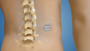
The new device for chronic back pain is implantable spinal neurostimulator. It stands as a remarkable innovation in treating chronic back pain, offering an advanced solution for managing persistent pain that other methods or conventional treatments cannot effectively control. This device offers hope in treatment for back pain sufferers who didn’t find relief with standard treatments.

The implantable spinal neurostimulator uses an under-skin electrical pulse generator, along with electrodes near the spinal cord or affected nerves. These electrodes emit controlled electrical impulses, which intervene in the transmission of pain signals to the brain, allowing the patient to control or reduce pain sensations.

Customizable electrical stimulation in this device adapts to each patient’s needs, granting better symptom control with therapy adjustments. This aspect allows for adaptability of therapy according to fluctuations in pain and the specific needs of the patient, thus providing greater control over symptoms.

The implantable spinal neurostimulator has evolved significantly in terms of its technology and features. Modern devices include improved sensors, personalized therapy, and mobile compatibility for easier patient monitoring and stimulation adjustment.

Treatment of Back Pain: Innovative Methods in Modern Medicine


Currently recommended treatment for back pain

Recommended treatment for back pain can involve a number of approaches, starting with lifestyle changes, physical therapy and specific exercises to strengthen the back muscles. Physicians may prescribe medications like non-steroidal anti-inflammatory drugs (NSAIDs) or pain relievers for pain management. In certain scenarios, complementary therapies such as acupuncture or therapeutic massage might also come under consideration.

Non-invasive approaches:

1. Lifestyle changes and conservative therapies

Exercise and physical therapy: Adapted exercise program can help strengthen back muscles, improve flexibility and reduce muscle tension.
Correct posture: Adopting correct posture can reduce pressure on the spine and prevent back pain.

2. Medications for pain management

Nonsteroidal anti-inflammatory drugs (NSAIDs): These drugs can reduce inflammation and pain associated with spinal conditions.
Analgesics: May be prescribed to reduce pain sensations, helping the patient manage the discomfort.

3. Complementary therapies

Acupuncture: Acupuncture points can be used to reduce pain and relax tense muscles.
Therapeutic massage: Massage can help relax muscles and relieve discomfort associated with back pain.

Invasive or interventional options:

1. Injections

Epidural injections: May be used to administer anti-inflammatory drugs directly near the affected spinal nerves to reduce inflammation and pain.

2. Surgical therapies

Spinal surgery: Recommended for severe cases or when other methods fail, addressing spine structural issues.

Treatment of Back Pain Innovative Methods in Modern Medicine


The most powerful medicine for back pain

There is no single “strongest” medication for back pain, as effectiveness can vary from person to person. The effectiveness of a particular drug may vary depending on the specific cause of the pain, its severity, and the individual response of each patient.

However, in many cases, people consider nonsteroidal anti-inflammatory drugs (NSAIDs) among the most effective for managing back pain. These drugs reduce inflammation and pain linked to spinal issues like herniated discs or osteoarthritis.

NSAIDs are available in various forms such as ibuprofen, naproxen, diclofenac and others. They work by blocking the body’s production of chemicals called prostaglandins, which are responsible for the sensation of pain and inflammation. By reducing these chemicals, NSAIDs can help reduce the inflammation and pain associated with spinal conditions.

It is important to note that the use of NSAIDs must be accompanied by special attention to the dose and duration of administration, since these drugs can have negative side effects on the stomach, kidneys or other organs, especially with long-term administration or high doses .

Consult a specialist before starting or continuing NSAIDs or any back pain medication for a proper evaluation, considering your health and other regular medications.

Treatment of Back Pain Innovative Methods in Modern Medicine


The safest pain medication for long-term use

At recommended doses and supervised by a doctor, NSAIDs are safe for long-term pain management.

Even though NSAIDs are generally considered safe, using them for an extended period necessitates caution and regular medical monitoring. Long-term NSAID use poses risks like stomach ulcers, bleeding, and organ damage, requiring supervised use with recommended doses.

To minimize the risks associated with long-term NSAIDs, it is crucial to follow your doctor’s directions and instructions regarding dosage and duration of treatment. Note any symptoms or side effects while using these meds; seek immediate medical attention for any issues or complications.

Long-term back pain management may involve safer alternatives: other pain relievers or non-drug therapies like physical therapy, acupuncture, or tailored exercise programs.

It is important to discuss any concerns or questions about long-term pain medication use with your doctor so that you can choose the safest and most effective treatment option tailored to your specific needs. Medical monitoring and supervision are essential to ensure safe and effective long-term back pain treatment.

The effects of Shockwave therapy in chronic back pain

The effects of Shockwave therapy in chronic back pain

Shockwave therapy, an innovative and promising method in the fight against chronic pain. It also has gained popularity and become the subject of increased interest in the medical field. This revolutionary technique has attracted attention for treating various conditions, including chronic back pain.

Shockwave therapy, with high-intensity acoustic waves, holds promise for relieving pain and enhancing function in those with debilitating back issues. Embraced by researchers and doctors, this innovative approach offers hope to those grappling with chronic back pain.


CONTENT:

  1. What is Shockwave therapy?
  2. How does Shockwave therapy work for back pain?
  3. The benefits of Shockwave therapy for back pain
  4. Conclusion


What is Shockwave therapy?

Shockwave therapy has evolved as a therapeutic solution in a wide range of conditions, from soft tissue injuries to musculoskeletal disorders and even chronic diseases. Medical practitioners widely adopt this therapeutic method for treating conditions such as herniated discs, chronic low back pain syndrome. The devices used for Shockwave therapy emit high-energy pulsed acoustic waves directed at the affected area of the patient’s back.

These shock waves act as stimuli that trigger biological reactions in the body. They cause the release of growth factors and other chemicals that contribute to tissue regeneration and repair. Thus, this therapy can help heal and strengthen the affected area, reducing inflammation and relieving pain.

Therapy boosts blood flow, aiding healing and restoring back function. Shockwave therapy aids chronic back pain by enhancing tissue regeneration, offering a non-invasive management option.

chronic back pain


How does Shockwave therapy work for back pain?

The mechanism by which Shockwave therapy works on back pain is a subject of ever-evolving research. However, there are several theories that try to explain how this form of treatment can relieve discomfort and improve the condition of patients.

Acoustic waves in this type of therapy stimulate tissue healing, aiding back pain treatment. Experts believe that shock waves trigger the release of growth factors and other chemicals in affected tissues, expediting the repair and rebuilding of damaged spinal tissues. Reducing inflammation, aiding healing, and improving back functionality are essential aspects.

Shockwave therapy may alter neural circuits, affecting pain perception and transmission to the brain. The influence on pain signal processing can reduce pain intensity, offering an analgesic effect and easing symptoms of chronic back pain.

Further research is necessary to fully understand the exact mechanisms by which this therapy operates in treating back pain. Preliminary results suggest significant benefits in managing chronic back pain, enhancing patients’ quality of life.


The benefits of Shockwave therapy for back pain

The benefits of this therapy in the treatment of chronic back pain are varied and have been recognized as significant factors in choosing this therapeutic method:

  • Pain reduction

Shockwave therapy has demonstrated the ability to reduce pain associated with conditions such as herniated discs or chronic low back pain syndrome. This reduction in discomfort can greatly improve patients’ quality of life, allowing them to engage in activities without being limited by persistent pain.

  • Improved mobility

Another major benefit of this type of therapy is improved back mobility and flexibility. Reducing inflammation and boosting tissue regeneration, therapy restores motion, aiding daily activities.

  • Non-invasive method

This therapy is non-invasive, which is a major advantage for patients. It does not involve surgical incisions or the introduction of substances into the body, thus reducing the risk of complications associated with other procedures. This feature makes the therapy an attractive option for those who want to avoid surgery or long-term drug treatments.

  • Fast recovery

Another important aspect is that Shockwave therapy can help patients recover faster. By stimulating the body’s natural healing processes, this form of treatment can speed up the recovery process, allowing patients to return to their usual activities in a shorter amount of time. Especially helpful for a swift return to normal routines without lingering pain or movement restrictions.

Overall, the therapy benefits chronic back pain by reducing pain, improving mobility, and aiding quick recovery, offering patients a promising alternative in managing this debilitating condition.

The effects of Shockwave therapy in chronic back pain

Conclusion

Shockwave therapy is a promising therapeutic option for those suffering from chronic back pain. However, it’s crucial to consult a specialist before beginning any treatment, as each patient’s needs and responses may vary. This therapy may also be used as part of a larger treatment plan that may include exercise, physical therapy, and other pain management methods.

In conclusion, this therapy offers relief and improvement for chronic back pain, providing hope to restore patients’ quality of life without invasive methods or medication risks.

Coxa Vara Understanding the Causes, Diagnosis, and Therapeutic Options for Hip Deformity

Coxa Vara: Understanding the Causes

Coxa Vara is an orthopedic condition in which the oblique angle of the femoral head (upper part of the thigh bone) relative to the shaft of the thigh bones is less than normal. This anomaly causes hip joint displacement, impacting mobility and daily activities.

This condition can appear either during the child’s development or later in life. In children, Coxa Vara may be congenital or may appear during growth. Congenital refers to the fact that this condition can be present from birth, as a result of abnormal development of the hip bones during the embryonic period.

Some genetic abnormalities or disorders in the development of bone tissues can contribute to the appearance of Coxa Vara in children.

While common in children, Coxa Vara can also arise in adulthood due to bone or joint-affecting conditions like Blount’s disease or skeletal dysplasia.  Factors such as obesity or excessive stress on the hip joint due to strenuous physical activities can also contribute to the development of this condition in adults.


CONTENT:

  1. Causes of Coxa Vara
  2. Coxa Vara diagnosis
  3. Therapeutic Options
  4. Prognosis and Recovery
  5. Conclusion


Causes of Coxa Vara

Coxa Vara is a complex orthopedic condition that can be caused by several factors. Here is a more detailed development of the root causes:

1. Congenital factors:

Abnormal development of the hip bones during the embryonic period is considered one of the possible causes of this condition. Some cases stem from genetic abnormalities or disturbances during embryonic development, leading to a congenital form of Coxa Vara with misaligned femoral head and thigh bone shaft angles, deforming the hip joint structure from birth.

2. Injuries or fractures:

Childhood trauma and fractures can directly affect the hip bones and later development of the femoral head. Fractures or severe injuries to the hip or femur can affect the normal growth and development of the bone, leading to changes in the angle of the femoral head relative to the shaft of the femur. These changes can contribute to the appearance of Coxa Vara during the child’s growth and development.

3. Diseases and medical conditions:

There are also certain medical conditions or diseases that can influence bone development and structure, contributing to Coxa Vara. For example, Blount’s disease impacts the tibia and may affect femoral head development. Skeletal dysplasia leads to bone tissue issues, contributing to hip joint deformities like Coxa Vara.


Coxa Vara diagnosis

This condition is diagnosed by an orthopedic doctor or a pediatric orthopedic specialist through multiple steps to confirm its presence in a patient. Here is a more detailed development of the diagnostic process:


Physical examination

The doctor will perform a detailed physical examination to evaluate the function and mobility of the hip joint. This assessment may include:

  • Observing the patient’s gait and weight bearing on the affected legs.
  • Testing the range of motion of the hip joint to note any restrictions or difficulty in movement.
  • Palpation and direct assessment of the hip area to detect possible tender areas or anatomical abnormalities.

Medical imaging

For Coxa Vara diagnosis and severity assessment, imaging tests like:

  • X-rays: These provide detailed images of the hip bones and can highlight structural changes in the femoral head and shaft of the thigh bones. Radiographs can help measure the abnormal angle of the femoral head in relation to the thigh bone, providing essential information for diagnosis.
  • MRI or CT: Magnetic resonance imaging (MRI) or computed tomography (CT) can provide more detailed and accurate images of the bone structure and soft tissues around the hip joint. These techniques can be useful to assess anatomic changes and provide a broader perspective on the condition.

Coxa Vara Understanding the Causes

Therapeutic Options

Treatment options for this condition depend largely on the severity of the condition and its impact on hip joint function. In general, treatment can range from non-surgical options for mild to moderate cases to surgery for severe or progressive cases.


Non-surgical treatment

  • Physical therapy and specific exercises: Physical therapy and tailored exercises may aid mild to moderate Coxa Vara, strengthening hip muscles and enhancing mobility. These exercises are designed to increase flexibility, improve muscle strength, and correct muscle imbalances that may be contributing to Coxa Vara symptoms.
  • Assistive devices: Sometimes the use of assistive devices such as crutches or braces can help reduce pressure on the hip joint and relieve discomfort during movement.

Surgery

  • Osteotomy: Surgery may be needed for severe cases to correct femoral head malalignment or thigh bone alignment. Osteotomy corrects femoral head angle by controlled bone cuts.
  • Hip reconstruction: In some severe cases, where Coxa Vara is associated with other hip joint conditions, joint reconstruction surgery may be necessary to restore the function and stability of the joint.

Coxa Vara Understanding the Causes

Prognosis and Recovery

The outlook and recovery for Coxa Vara patients varies depending on the severity of the condition and the treatment applied. In the case of surgery, the recovery period may require extensive physical therapy to restore strength and mobility in the hip joint.


Conclusion

This orthopedic condition can affect the quality of life and functionality of the hip joint. Early diagnosis and appropriate treatment are crucial to managing this condition and preventing long-term complications. Seek evaluation by an orthopedic specialist for personalized treatment guidance.

Fonduri de investiții – 4 aspecte generale pe care trebuie să le cunoști

Jade therapy for back pain

Jade therapy is an extraordinary solution even for those who suffer from advanced disc herniations when the pain goes down on the leg as well as for those in sciatica or lumbago crisis. It is the most performing system globally and offers a profound therapeutic experience. From a therapeutic point of view, it combines a very wide range of principles:

  • Acupressure
  • Thermotherapy
  • Mechanical elongation of the spine
  • Massage

Back pain is pain in any region of the back. It is divided into cervical, thoracic back pain, lower back pain (lumbar) or sacral pain. Additionally, the lumbar region is the most prevalent area for pain as it primarily supports the weight of the upper body.

Episodes of back pain can be acute, sub-acute, or chronic, depending on the duration. Moreover, back pain is one of the most common reasons why people go to the doctor and one of the leading causes of disability worldwide. Most people have back pain at least once in their lifetime.

Jade bed therapy does not require any effort on the part of the patients. A session with this device lasts 45 minutes. Furthermore, a package of 10-12 sessions is recommended for long-term results. Additionally, it is mandatory to consume 250-400 ml of water before the procedure and 250-400 ml of water immediately after the procedure.

The only contraindication for this device is that there is no metal rod implant in the spine.


CONTENT:

  1. How does jade therapy work for back pain?
  2. How an automated Jade massage bed works
  3. Combining elongation procedures with spine toning procedures
  4. Benefits of Jade Therapy
  5. Warning


How does jade therapy work for back pain?

Jade therapy for back pain is based on the principles of heat and pressure therapy. Additionally, this can be done by various methods:

  • Jade stone massage: The therapist may use smooth, pre-heated jade stones to massage painful areas of the back. Furthermore, the heat from jade stones relaxes tense muscles and can help reduce inflammation and discomfort.
  • Jade wraps or compresses: Jade stones can be heated and placed on the affected area or wrapped in cloth to create compresses applied directly to the back. Additionally, this process can help warm the tissues, relax the muscles, and relieve pain.
  • Jade Therapy Pads: There are devices that use jade therapy pads that can be applied to the concerned back. They are designed to emit heat and relax painful areas.


How an automated Jade massage bed works

The action of the massage bed comes in two directions:

1. Mechanical elongation of the spine as a therapeutic principle

All 7 programs, by reducing the rolls of paravertebral jade, along the spine, according to the natural curvature of the spine, achieve mechanical elongation of the spine with the effect of decompressing the intervertebral discs and increasing the spaces between the vertebrae.

2. Long-term, infrared radiation therapy (Far-Infrared Technology) as a therapeutic principle

Throughout the 7 massage programs, they heat the jade rolls, either based on your personal choice or as per the specialist’s recommendation, to temperatures between 50 and 70 degrees Celsius.

All programs use infrared thermotherapy (heat) and all programs stimulate and improve blood circulation, calming and completely eliminating the symptoms of poor circulation, contributing to a strong detoxification on all 5 pathways, relaxes muscles, eliminates inflammation in the body and contributes strongly to pain therapy, eliminating it.

Combining elongation procedures with spine toning procedures

Through the automated massage bed with Jade therapy, we obtain the elongation of the spine, decompression, and repositioning of the vertebrae. Now, we should ask ourselves why the spine got into the situation where it needed mechanical intervention to eliminate back pain.

In most cases, sedentary lifestyle, overweight and atrophy of the back muscles are the causes that generate these episodes of back pain.

Given that most of you lead a sedentary lifestyle and your daily routine lacks physical activity, heading to the gym may not be the ideal solution for spinal health. Engaging in weightlifting exerts pressure on the spine, potentially causing vertebral compression over time. Furthermore, improperly executed exercises can have lasting adverse effects on the spine.


Benefits of Jade Therapy

  • Deep muscle relaxation
  • Stimulating and improving blood circulation
  • Better oxygenation of muscles, tissues, brain
  • Elimination of insomnia
  • Elimination of emotional problems: depression, panic attacks, anxiety
  • Elimination of BURN OUT syndrome – chronic fatigue, exhaustion, stress
  • Elimination of headaches
  • Elimination of menstrual pain
  • Elimination of back pain
  • Hot spine elongation
  • Elimination of joint pain
  • Elimination of rheumatic pain
  • Deep detoxification
  • Elimination of chronic constipation
  • Hormonal balance

Warning

Even though jade therapy may help some individuals with back pain, it’s crucial to talk to a doctor or therapist before trying any alternative treatment. Also, make sure it’s safe for your health and avoid using jade stones on irritated or damaged skin.

Disc protrusion symptoms, diagnosis and treatment

Disc protrusion: symptoms, diagnosis and treatment

Disc protrusion is a common spinal condition that can cause pain and discomfort for those affected. To better understand this problem, it is important to examine the anatomy of the spine and understand what exactly constitutes a disc protrusion.

The spine comprises a series of bones known as vertebrae, which are separated by intervertebral discs. These discs consist of a sturdy outer ring called the annulus fibrosus and an inner gelatinous substance known as the nucleus pulposus. They serve as shock absorbers and allow fluid movement of the spine.

Disc protrusion occurs when the gelatinous inner part of the intervertebral disc begins to protrude from the annulus fibrosus. This can be caused by various reasons, such as trauma, the natural wear and tear of the disc over time, or an incorrect body position that puts pressure on the disc.


CONTENT:

  1. Symptoms of Disc protrusion
  2. Diagnosis of Disc protrusion
  3. Treatment for Disc protrusion


Symptoms of Disc protrusion

The symptoms of a herniated disc are diverse and can vary considerably depending on the location and size of the herniation, as well as the level of pressure exerted on nerves or other surrounding structures. These symptoms can affect a person’s quality of life and ability to carry out normal daily activities. Here are some common symptoms of a herniated disc:

1. Localized and radiating pain

Pain is a characteristic symptom of a disc protrusion. This sensation can manifest as sharp, dull, persistent pain, or discomfort in the affected spinal area. The pain may radiate to other parts of the body, such as the legs or arms, depending on the level of the hernia and the affected nerves. For example, a lumbar disc herniation can cause pain that radiates down one or both legs, known as sciatica.

2. Muscle weakness and abnormal sensations

The pressure exerted by the herniated disc on the nerves can cause abnormal sensations such as tingling, numbness or burning sensations in the affected area. Muscle weakness in the area may also occur. For example, in the case of a cervical disc protrusion, these abnormal sensations can be felt in the arms and hands.

Disc protrusion symptoms, diagnosis and treatment


3. Limitation of mobility

A disc protrusion can affect the normal mobility and flexibility of the spine or surrounding regions. Affected individuals may experience difficulty performing specific movements, such as bending or turning the body, without experiencing pain or discomfort.

These symptoms can vary in intensity and can appear suddenly or develop gradually over time. It is important to pay attention to early signs and seek medical attention if symptoms persist or worsen. Early diagnosis and treatment can help manage symptoms and prevent long-term complications associated with a herniated disc. A doctor or health professional can recommend additional tests and an appropriate treatment plan to effectively manage this condition.


Diagnosis of Disc protrusion

Diagnosing a herniated disc involves a series of steps and investigations performed by doctors or health professionals to identify and confirm the presence of this spinal condition. Disc protrusion can be diagnosed after a careful evaluation of symptoms, physical examination, and imaging investigations. Here’s how the diagnostic process typically works:

  • Medical history and physical exam

The doctor will take a detailed history to understand the patient’s symptoms, how they have evolved and how they affect quality of life. A comprehensive physical examination will evaluate mobility, muscle strength, reflexes, and sensation in the affected areas.

  • Assessment of symptoms

The diagnostic process takes into account common symptoms such as localized or radiating pain, muscle weakness, and abnormal sensations. Details regarding the duration, intensity, and how the symptoms affect daily activities are essential for the physician in formulating an accurate diagnosis.

  • Imaging investigations

Medical imaging is essential in confirming the diagnosis of disc protrusion. These investigations may include:

X-rays: Can give a picture of the bone structure and rule out other conditions of the spine, but do not give precise details about the intervertebral discs.

MRI (magnetic resonance imaging) or CT (computed tomography): These imaging methods provide clearer and more precise details of the intervertebral discs, nerves and surrounding tissues. MRI is more commonly used to evaluate the disc and nerves, providing detailed images in different planes of the spine.

  • Other additional tests

Occasionally, healthcare professionals might require additional tests like electromyography (EMG) or nerve conduction tests to assess nerve function and ascertain if the herniated disc is exerting pressure or impacting the nerves.

Correct diagnosis of disc protrusion is crucial to develop an appropriate treatment plan. After confirming the presence of a herniated disc, the doctor might suggest various treatment options, including physical therapy, medications, injections, or, in severe cases, surgery. It is important to seek specialist medical attention for the evaluation and proper management of disc protrusion, given that each patient may require a personalized approach depending on their clinical characteristics and the severity of the condition.

Disc protrusion symptoms, diagnosis and treatment

Treatment of Disc protrusion

Treatment for disc protrusion is variable and may involve a combination of conservative methods and medical or surgical interventions, depending on the severity and location of the problem.

Here are some of the treatment options for a herniated disc:

Rest and activity modifications

Relative rest and avoidance of activities that aggravate pain are often recommended initially. Maintaining proper posture and avoiding movements or positions that put pressure on the spine can help reduce symptoms.

Physical therapy

Exercise and movement therapy are essential in the management of disc protrusion. The physical therapy program may include back strengthening exercises, stretching, and correcting posture to reduce pressure on the affected disc.

Medications

Healthcare providers may prescribe nonsteroidal anti-inflammatory drugs (NSAIDs) like ibuprofen or naproxen to reduce inflammation and pain. They might also recommend pain relievers to manage discomfort. Muscle relaxants could be prescribed in some cases to alleviate muscle spasms associated with a herniated disc.

Alternative therapies

Some people find benefits in complementary therapies such as acupuncture, chiropractic or massage therapy. These treatments can help relax the muscles and relieve the pain associated with the herniated disc.

Epidural injections

Epidural corticosteroid injections may help reduce inflammation and pain in the affected area by administering them near the affected disc or irritated nerve.

Surgery

In severe cases or when conservative treatments do not bring relief, surgery may be an option. Surgery may involve removing the part of the bulging disc that is putting pressure on the nerve or other surrounding structures. Surgery can be performed through various procedures such as discectomy, laminectomy or microdiscectomy.

It is important to note that each case of disc protrusion is unique and requires an individualized approach to treatment. Before starting any treatment program, it is essential to consult a doctor or health professional to obtain an accurate diagnosis and develop an appropriate treatment plan.

Also, adopting a healthy lifestyle that includes regular exercise, maintaining a healthy weight, and avoiding positions or movements that can strain the spine can help prevent or reduce the risk of a herniated disc.

Cervical Massage: Indications, Benefits and Procedures

Cervical Massage: Indications, Benefits and Procedures

Cervical massage is an effective and relaxing therapeutic technique aimed at the neck region and the cervical area of the spine. This form of massage is used to relieve muscle tension, reduce pain, and improve mobility and flexibility in the neck and shoulders.

Indications for cervical massage are varied and include various conditions and situations that can benefit from this therapy.

CONTENT:

  1. Indications and Situations That Benefit from Cervical Massage
  2. The benefits of Cervical Massage
  3. The Cervical Massage Procedure: A Personalized Experience for Neck and Shoulder Health

Curious about how cervical massage can relieve neck and shoulder tension but short on time? Watch this quick video to learn the key benefits, techniques, and tips for maximizing its effectiveness. Or, read on for a detailed breakdown!

Indications and Situations That Benefit from Cervical Massage

The sedentary lifestyle, stress, and incorrect positions during work or daily activities can cause various discomforts and cervical dysfunctions. Cervical massage emerges as a holistic remedy offering multiple benefits:

  • Tension and Muscle Spasms

A common cause of neck pain and stiffness in this area is muscle tension and spasms. Cervical massage is extremely effective in relaxing tense muscles and reducing spasms, helping to relieve discomfort.

  • Neck Pain and Stiffness

People who experience chronic neck pain or a constant feeling of stiffness in the cervical area can benefit from massage therapy to reduce pain and improve neck mobility and flexibility.

  • Migraines and Headaches

Some migraines or headaches may be related to tension in the neck and shoulder area. By relaxing tense muscles and tissues, cervical massage can help reduce the frequency and intensity of these pains.


  • Recovery from Trauma or Accidents

Cervical massage can be part of the recovery process for those who suffer from neck trauma or injuries, whether from accidents or sports activities.

The benefits of Cervical Massage

Cervical massage, an established form of manual therapy, brings many benefits to both the physical health and the mental state of the individual. These benefits are supported by multiple therapeutic effects aimed at relieving discomfort and strengthening neck and shoulder health:

  • Reducing Muscle Tension and Improving Mobility

Moreover, tense and strained muscles in the cervical area are prone to developing muscle knots, leading to pain and restricted movement. However, a qualified therapist administering cervical massage can effectively relax and release these tense regions, diminishing discomfort and notably enhancing neck flexibility and mobility. Employing strategic massage techniques on these problematic areas serves to loosen muscle knots and reinstate regular muscle tone.

  • Relief of Discomfort and Pain

Through specific techniques, cervical massage can alleviate and reduce the discomfort associated with neck pain, tense shoulders or stiffness in this region. By gently manipulating tense muscles and tissues, massage helps to relax them and reduce feelings of discomfort and pain. Also, stimulation of nerve receptors can trigger the release of endorphins, natural neurotransmitters that can help reduce the sensation of pain.

  • Improving Blood Circulation and Tissue Oxygenation

Another major benefit of cervical massage is the stimulation of local blood circulation. Applying pressure and precise movements supports blood flow, enhancing oxygenation and nutrient supply to tissues. Improved blood circulation aids tissue healing, expediting recovery from minor injuries or muscle strains.

  • Relaxation and Stress Reduction

Cervical massage is recognized for its ability to induce a deep state of relaxation. Through gentle and rhythmic movements, the therapist can help reduce stress and anxiety levels. This relaxing experience can have a positive effect on the whole body and mind, contributing to a general state of well-being and inner calm.


  • Support in Maintaining Correct Posture

In addition to the immediate benefits, cervical massage therapists can provide advice and exercises to help maintain proper posture. Proper posture is essential for long-term neck and shoulder health, and these recommendations can help prevent the recurrence of cervical discomfort.

The Cervical Massage Procedure: A Personalized Experience for Neck and Shoulder Health

This type of massage is not just a simple application of pressure and movements in the area of the neck and shoulders, but represents a personalized therapeutic session, adapted to the specific needs of each individual. Here’s how a typical massage session goes in detail:

1. Preliminary Assessment

Each massage session begins with an essential stage – the initial assessment of the condition of the neck and shoulders. The therapist will talk with the client to better understand the specific issues they are facing. This discussion may include your medical history, level of pain or discomfort, problem areas, and any other relevant issues.

After the initial dialogue, the therapist will perform a detailed physical assessment. Examining the cervical area and shoulders to identify tension, muscle knots, stiffness, or any physical issue. This initial assessment is crucial to tailoring the massage session to suit each individual’s specific needs.

2. Adaptation of the Massage Session

Based on the initial assessment, the therapist will tailor the massage session to the client’s individual needs and preferences. Each person has different levels of sensitivity, pressure tolerance and preferences regarding massage techniques.

The therapist will constantly communicate with the client to ensure that the pressure and intensity applied is comfortable and beneficial. The therapist employs different massage techniques like light/firm pressure, circular/elongated movements, or kneading to relax tense muscles in the cervical area.

3. Using Massage Techniques

The therapist will use various techniques and specific movements to work on the areas identified during the assessment. These techniques may include friction, light or deep pressure, gentle stretching, and deep massage to release tension and relax tight muscles.

The therapist might use oils or creams for smoother hand movement and to avoid excessive skin friction.

Cervical Massage Indications, Benefits and Procedures


4. Completion and Recommendations

At the end of the massage session, the therapist will provide recommendations to maximize the benefits of the therapy. These may include stretching exercises, suggestions for improving posture, or other tips to maintain relaxation and prevent recurrence of muscle tension.


In conclusion, this form of massage stands as a valuable therapeutic option for individuals experiencing discomfort and tension in the neck and shoulder area. Through the relaxation of tense muscles, enhancement of blood circulation, and stress reduction, cervical massage significantly contributes to overall health and quality of life. Therefore, medical referral and collaboration with specialized therapists play a pivotal role in achieving optimal results.

Diagnosis of back pain

Diagnosis of back pain

In most cases, a specialist is able to make the diagnosis after physical examination and medical history, and imaging tests are done only to confirm the diagnosis and provide additional information.

Low back pain is a common problem, especially in developed countries. These pains can have several causes, so it is recommended to consult a specialist to determine the exact cause of the condition that leads to back pain.

After low back pain, it is advisable to go to the family doctor / general practitioner. He will perform a physical examination and discuss his symptoms and medical history with his patient, and then determine which doctor will refer the patient to.

The diagnosis for a disease presenting symptoms of low back pain can be made by a variety of specialists, including orthopedists, rheumatologists, neurologists, or even internists if there is suspicion that the pain may be related to an organ disease radiating to the lumbar area elsewhere (for example, kidney disease).

A complete medical history and physical examination can usually identify any serious condition that may be causing the pain. During the exam, the doctor will ask about the onset, location, and severity of the pain; duration of symptoms and any limitations in movement; history of previous episodes or any health conditions that may be related to pain.

Along with a detailed examination of the back, neurological tests are performed to determine the cause of the pain and the appropriate treatment. The cause of chronic lower back pain is often difficult to determine even after a thorough examination.

Imaging tests are not justified in most cases. Under certain circumstances, healthcare providers may recommend imaging to rule out specific causes of pain, including tumors and spinal stenosis.


CONTENT:

  1. Magnetic resonance imaging (MRI)
  2. Ultrasound imaging
  3. Bone scans
  4. Blood tests
  5. Radiography
  6. Discography
  7. Computed tomography (CT)
  8. Myelograms


Magnetic resonance imaging (MRI)

It uses a magnetic force instead of radiation to create a computer generated image. Unlike X-rays, which show only bone structures, MRI scans also produce images of soft tissues such as muscles, ligaments, tendons, and blood vessels.

An MRI may be recommended if a problem such as infection, tumor, inflammation, herniated disc, rupture, or nerve compression is suspected. MRI is a non-invasive way to identify a condition that requires prompt surgical treatment. However, in most cases, an MRI scan is not required during the early stages of back pain.


Ultrasound imaging

They are also known as ultrasound scanning or sonography, which utilizes high-frequency sound waves to obtain images inside the body.  Sound wave echoes are recorded and displayed as real-time visual images. Ultrasound imaging can show tears in ligaments, muscles, tendons and other soft tissue masses in the back.


Bone scans

They are used for detecting and monitoring infections, fractures, or bone disorders. A small amount of radioactive material injected into the blood collects in the bones, especially in areas with an abnormality. The images generated by the scanner can identify certain areas of irregular bone metabolism or abnormal blood flow, as well as measure levels of joint disease.


Blood tests

These are not commonly used for diagnosing the cause of back pain. However, in some cases, healthcare providers may perform them to search for indications of inflammation, infection, or the presence of arthritis. Potential tests include erythrocyte sedimentation rate and C-reactive protein.


Radiography

It is often the first imaging technique used to look for broken bones or injured vertebrae. X-rays show bone structure and any vertebral misalignment or fractures. Soft tissues, such as muscles, ligaments, or clogged discs, are not visible in conventional X-rays.


Discography

It can be used when other diagnostic procedures fail to identify the cause of the pain. This procedure involves injecting a contrast dye into a spinal disc suspected to cause low back pain. Fluid pressure on the disc will reproduce the person’s symptoms if that disc is the cause.

The dye will show damaged areas on CT scans performed after injection. Discography can provide useful information in cases where people are considering lumbar surgery or where their pain has not responded to conventional treatments.


Computed tomography (CT)

It visualizes structures of the spine that an X-ray cannot capture, such as a ruptured disc, spinal stenosis, or tumors. Using a computer, CT scanning creates a three-dimensional image from a series of two-dimensional images.


Myelograms

They increase the diagnosis of X-ray and CT images. In this procedure, the doctor injects a contrast dye into the spinal canal to enable visualization of compression of the spinal cord and nerves, caused by herniated discs or fractures, on an X-ray or computerized tomography (CT) scan.

Long-term effects of COVID-19 infection

Long-term effects of COVID-19 infection

COVID-19 infection is proving to be much more than a respiratory illness. It can affect many organs, not just the lungs – from the skin and muscles to the eyes, heart and kidneys – creating long-term health problems. These include fatigue, blurred vision, difficulty breathing, muscle aches, confusion, headaches and even hallucinations.

Most people infected with the new coronavirus tend to have mild to moderate forms of the disease and heal within a few weeks. In contrast, people who survive COVID-19 infection after needing intubation and longer hospitalization may be left with a number of long-term side effects.


CONTENT:

  1. Long-term effects of COVID-19 infection known to date
  2. Long-term side effects – a feature of other viral infections


Long-term effects of COVID-19 infection known to date

Although there is still much to learn from people who have recovered from COVID-19, there are a number of long-term effects that doctors have seen so far.

Lung problems

We know that COVID-19 infection primarily attacks the lungs, causing inflammation. For this reason, survivors may experience long-term breathing difficulties. Some people recovering from COVID-19 may experience a dry cough or pain when breathing, and patients who have needed mechanical ventilation may have more severe symptoms.

Along with the lung pain, there is discomfort in the back area. You shouldn’t be surprised that once you get in touch with the virus, your back starts to hurt.


Neurological problems

Research shows that COVID-19 infection can affect the brain and central nervous system. Some people who have recovered have reported symptoms such as headaches, dizziness, difficulty concentrating or memory, and even hallucinations. Researchers say that these symptoms are more common in patients who have had severe forms of the disease.


Heart problems

A study by Frankford University in Germany showed abnormal heart rates in more than 75% of those studied who recovered from COVID-19. It was found that a considerable majority of patients in the study had inflammation of the heart and mucous membranes. This can be disturbing, as inflammation of the heart can lead to long-term heart disease.

As we are dealing with a relatively new disease, it is not yet known why certain patients experience one or more side effects after infection with the new coronavirus, how long these effects persist and whether COVID-19 infection will cause certain chronic diseases. , who will need lifelong monitoring.


Long-term side effects – a feature of other viral infections

Long-term symptoms of a severe viral illness are not new. Infections with other pathogens are associated with long-lasting symptoms, from heart problems to chronic fatigue. Even common illnesses, such as the flu and pneumonia, can lead to a long recovery. For example, the flu has long been linked to persistent symptoms, such as fatigue and muscle aches.

Infections with certain bacteria and Zika virus, among others, are linked to Guillain-Barre syndrome, a condition in which the immune system attacks the nervous tissue, causing muscle weakness and paralysis.

There is a precedent for infections with other coronaviruses. For example, the first SARS coronavirus and the Middle East Respiratory Virus (MERS) caused serious illness, and a significant number of patients developed acute respiratory disease syndrome (ARDS) and needed intensive care.

Canadian researchers found common issues among SARS survivors in Toronto: sleep disorders, fatigue, depression, and muscle aches. One-third of survivors were forced to change jobs and lifestyles, and only 14% had no long-term symptoms. A Korean study found that 48% of MERS survivors experienced chronic fatigue within a year of recovery.

Many severe COVID-19 patients require a long recovery period, as widely recognized in the medical community. The virus can attack the heart through direct invasion and inflammation, damaging or destroying heart cells and affecting function. Also, the virus disrupts ACE2 receptors, which protect heart cells and degrade angiotensin II, a blood pressure-raising hormone.

Back pain caused by Omicron

Back pain caused by Omicron

Doctors have reported an increase in patients’ back and muscle pain complaints since the onset of the Omicron wave, and data from various countries confirm this.

Since the onset of the coronavirus pandemic, there has been a clear list of symptoms, from fever to difficulty breathing to loss of taste and smell. As the pandemic developed and COVID-19 variants multiplied, the list of symptoms grew.


CONTENT:

  1. Injuries to soft tissues and joints
  2. Why pain occurs
  3. Back pain, the newest symptom of the Omicron variant


Injuries to soft tissues and joints

With COVID-19, the pain complaints of many patients focus on the soft tissues around the joints. Those who have previously had back pain or joint problems may notice an occurrence of these problems after receiving Omicron.

The good news is that for the most part, back and muscle aches decrease and go away on their own when you recover, and the infection disappears from your body, which lasts about two weeks.

Because Omicron is still considered a new strain, there is not enough data to know if back pain will become one of its persistent symptoms. However, if you suffer from persistent back pain that does not improve over time, consult a doctor, whether or not you have had COVID-19.


Why pain occurs

A February report by the World Health Organization (WHO) looked at nearly 56,000 cases of COVID-19 in China and found that almost 15% of patients had muscle pain.
When you get sick, your body triggers an immune response against aggressors, such as harmful bacteria or viruses. Back and leg joint pain is due to the body’s inflammatory response to the virus.

These viral infections cause tremors, chills, body aches and difficulty moving.

This form of back pain feels like cramps or spasms in the back muscles.

Specifically, muscle aches and pains are a result of interleukin-releasing immune cells, which are proteins that help fight invasive pathogens.

Therefore, back pain may be in addition to the usual symptoms of COVID infection, such as fever, dry cough, loss of taste or smell, sore throat, headache or pain in other areas.


Back pain, the newest symptom of the Omicron variant

There are now reports of back pain, a new symptom of the Omicron variant. According to the Jerusalem Post, South Africa, which first reported Omicron, has seen an increase in reports of muscle aches, and especially back pain, among people infected with the virus. Indian and Norwegian data support this claim.

The cause of back pain is probably an increase in the level of inflammation in the body, as the immune system fights the invading virus, which stimulates the muscles and joints. Although it is still too early to know, some doctors suspect that Omicron has a unique effect on the bone and muscle system that is manifested by muscle pain.

While facing a viral infection, the body releases a lot of small proteins called cytokines that activate the immune system against pathogens. Cytokines can stimulate muscles and joints.

However, this virus is not the only one that causes this reaction. Back and muscle pain is a familiar symptom of all sorts of other viral and bacterial infections. This immune response, which is the body’s natural response to a viral infection, can lead to various complaints from patients related to the musculoskeletal system.

Spinal Infections

Spinal Infections

There are spinal infections that can affect the intervertebral discs. When it invades the vertebral plates (the cartilaginous structures that separate the intervertebral disc from the adjacent bone structure) and the vertebral bodies. they are called vertebral osteomyelitis.

If at the time of diagnosis the infection has already compromised 2 structures, it refers to spondylodiscitis. Moreover, this makes the purulent secretion can reach the epidural space (the space around the epidural sac – it is like a sac of tissue, made of dura mater), which causes the spinal cord/its last nerve fibers), causing a spinal epidural abscess.


CONTENT:

  1. What are the causes and factors that predispose to spinal infections?
  2. Symptoms
  3. Evolution and prognosis
  4. Diagnostic
  5. Treatment


What are the causes and factors that predispose to spinal infections?

  • Diabetes
  • Immunocompromised patients (also, organ transplant patients who are being treated with immunosuppressive drugs and intravenous drug users)
  • Alcohol abuse
  • Background surgery on the spine
  • Neoplastic diseases (cancer)
  • Rheumatic diseases
  • Liver cirrhosis
  • Renal insufficiency
  • Minimally invasive non-surgical epidural procedures


Symptoms

At the clinical examination:

  • Additionally, in children, loss of lumbar lordosis, fever, and neurological signs (muscle weakness, paresis, gait disorders) are most common.
  • Adults report local pain in the back – neck / neck, associated with fever and may have neurological deficits – muscle weakness in the legs, urinary incontinence
  • There is a clinical triad for spinal epidural abscess
    1. Back pain
    2. Fever
    3. Neurological deficits (muscle weakness, paresis, sphincter disorders – urinary retention)
  • Paravertebral muscle contracture
  • Root pain (pain that has the path of a nerve)
  • Stiff neck (patient cannot bring chin to chest)


Evolution and prognosis

The unfavorable prognosis in cases of spinal cord infection is associated with:

  • The advanced age of the patient
  • The infection is located in the cervical / thoracic segment of the spine
  • The presence of diabetes
  • Heart disease
  • Neurological disorders (muscle weakness, paresis) that have occurred for more than 36 hours
  • Late diagnosis
  • Infections with bacteria resistant to usual antibiotic treatment


Diagnostic

  • With the help of inflammatory markers analyzed in the blood collected from the patient:
    1. Red blood cell sedimentation rate (ESR)
    2. C-reactive protein level (PCR)
    3. The number of leukocytes
  • Bacteriological cultures of blood and urine collected from the patient
  • Wound culture that has clinical signs of infection
  • Imagistic:
    1. Standard radiography: fast, easy to perform, but has the disadvantage that it does not provide clear evidence in the case of an isolated epidural abscess and does not reveal information about an osteomyelitis in the early stages.
    2. Computed tomography (CT): highlights bone changes caused by an infection, pathological calcifications suggestive of bone tuberculosis.
    3. Contrast-enhanced nuclear magnetic resonance imaging (MRI): Moreover, is the investigation of electives, having a great ability to provide information about the local anatomy, the extent of infection, destruction of adjacent tissues, and possible compression of the spinal cord.


Treatment

NON-SURGICAL TREATMENT

The indication for surgery in patients with spinal infection and neurological impairment (muscle weakness, paresis, sphincter disorders) is not questioned, but there are controversies regarding the choice of treatment for patients with minimal or even without neurological changes.

You can opt for treatment with antibiotics, to which bacteria are sensitive—an aspect established after a previous antibiogram was performed—combined with external immobilization or surgery. Additionally, this depends on the neurological status of the patient at presentation, the severity of the disease, the extent of tissue destruction adjacent, and antibiotic response.

SURGICAL TREATMENT

Surgery is performed immediately if there are signs of compression on the spinal nerves or spinal cord. Moreover, the operation should be considered if there is local pain (back / neck) or in case of changes in the curves of the spine, after non-surgical treatment.

The purpose of surgery is:

  • Additionally, aggressive debridement of tissues affected by infection
  • Collection of biological material for bacteriological examinations
  • Decompression of spinal nerves and spinal cord
  • Drainage of purulent collections from neighboring structures of the spine