scoliosis

Scoliosis: causes, symptoms, treatment

Scoliosis is a deformity of the axis of the spine in the frontal, sagittal and horizontal planes. It mainly affects children and adolescents, but can also occur in adults, sometimes due to a disease or the existence of malformations. Scoliosis is twice as common in girls as in boys. It can be observed at any age but is much more common at the age of more than 10 years. It is hereditary, so people with scoliosis are very likely to pass it on to their children, in any case, there is no correlation between the severity of scoliosis from one generation to the next.

CONTENT:

  1. Causes
  2. Symptoms
  3. Treatment


What are the causes?

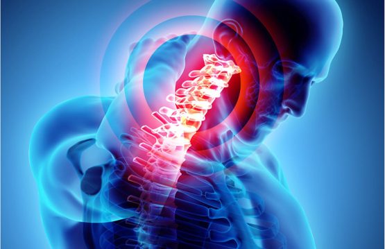
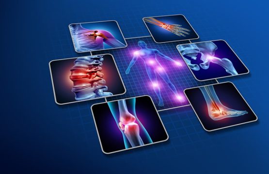
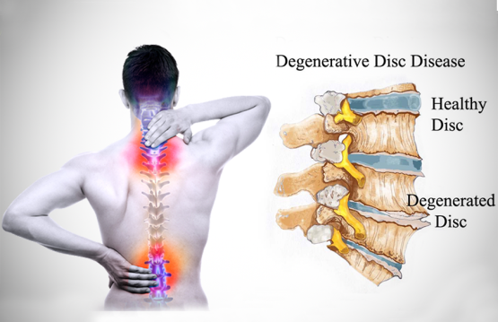
Scoliosis is a condition that leads to the curvature of the spine. It can affect any part of the spine, but especially the thoracic area (thoracic scoliosis) and the lower back (lumbar scoliosis). Most of the time, the causes of the change in the shape of the spine are not known, but doctors believe that it has to do with:

  • Some neuro-muscular disorders – these affect the muscles and nerves and include polio, muscular dystrophy and cerebral palsy;
  • Some genes – it is believed that there is at least one gene involved in the transmission of scoliosis;
  • Leg length – a person who has one leg longer than another can develop scoliosis;
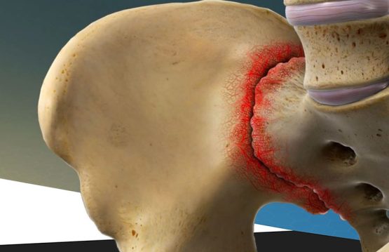
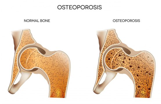
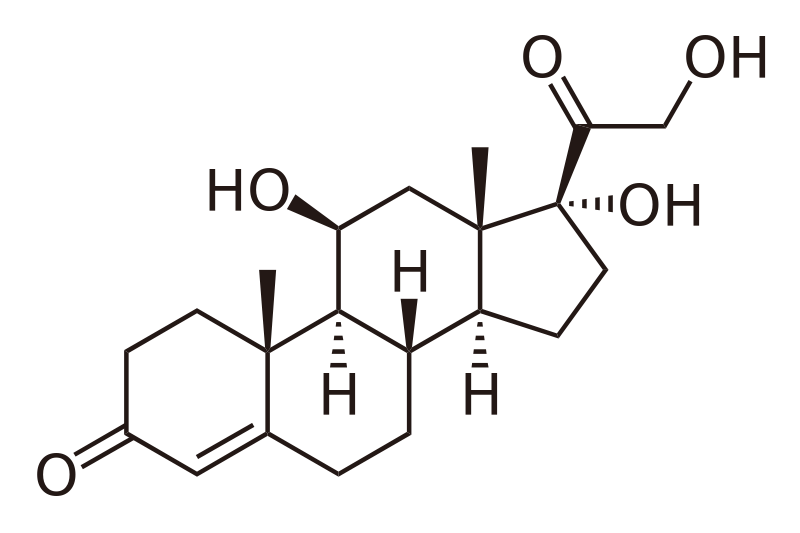
  • Osteoporosis – scoliosis can occur as an effect of this disease, once bone degeneration affects other areas of the body, so the spine;
  • Incorrect posture – at the office or inappropriate wearing of the backpack.


What are the symptoms?

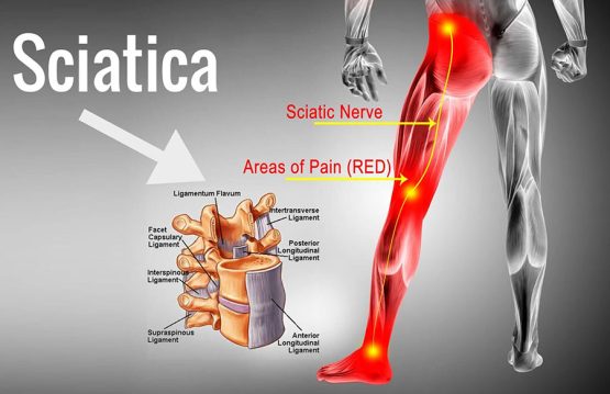
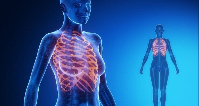
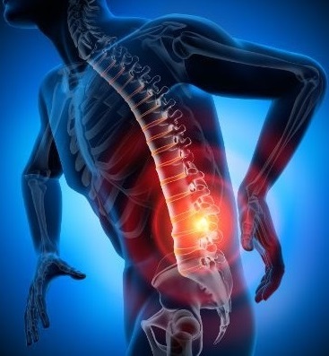
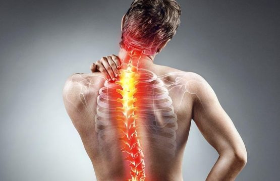
Often, the symptoms are noticed especially by those around. Once the spine grows, it pushes the shoulders and hips so that they can look asymmetrical. Most of the time, scoliosis does not affect movement and does not cause consistent back pain until the curvature becomes severe. Sometimes, unexplained back pain can occur because the curvature of the spine puts pressure on the spinal nerves and sometimes on the entire spinal cord. In addition to mild pain, at least at first, weakness, numbness, and pain in the lower extremities may occur. Another indication of scoliosis is the feeling of fatigue, after long periods of sitting or standing. As the spine becomes more deformed, the muscles must make an extra effort to keep the body aligned. Over time and in the absence of treatment, scoliosis progresses and puts pressure on the chest cavity, restricting the person’s ability to breathe easily and widely.  


What is the treatment?

Most adolescents who have a type of disease that does not have a clear cause are monitored at regular intervals (every 6 months). The corset The corset is the standard treatment for teenagers who have a spinal curvature between 25 and 40 degrees. The purpose is stiffening and temporary correction. Surgery Those who have a spine curve between 40 and 50 degrees should consider surgery. The doctor’s goal is to make sure that curvature does not get worse. However, the operation does not completely straighten the spine. During the procedure, metal implants are used to correct the curvature and to maintain a correct position until a bone graft is placed. Surgery for scoliosis involves joining the vertebrae, a process called spinal fusion. Many studies have shown that electrical stimulation and exercise programs cannot prevent the progression of scoliosis. However, people affected by this disease must remain active.

Sciatica

Sciatica: causes, symptoms and treatment

Sciatica is not a condition in itself, but a term we use to describe the manifestation of pain felt in the lower back in the lumbar region. From this point, the pain radiates to the hips and then descends to the leg. The pain is caused by compression of the sciatic nerve and usually occurs on one side of the body.

The sciatic nerve is the widest and longest nerve in the human body and consists of the nerve roots that start from the spinal cord (L5, which starts between the 4th and 5th lumbar vertebra and S1, which starts between the 5th lumbar vertebra and the first sacral vertebra) and descends to the ankles.

Sciatica can be of two types:

1. Acute, when it lasts between 4 and 8 weeks and can be treated with exercise, hot or cold compresses or painkillers.

2. Chronic, when it lasts more than 8 weeks and requires medical intervention (physiotherapy).

CONTENT: 

  1. Causes
  2. Symptoms
  3. Treatment


What are the causes of Sciatica?

Sciatica therefore means the pressure on the sciatic nerve. The causes can be multiple, but keep in mind that there is no obvious cause to explain the inflammation or irritation of the sciatic nerve. It can be about:

  • disc herniation: is the most common cause of sciatica (it is the movement of an intervertebral disc). The spine has 3 parts: the vertebrae (the bones that protect the afferent nerves), the nerves and the intervertebral discs. The discs between the vertebrae are made of cartilage, which acts as a shock absorber between the vertebrae and allows flexibility of the spine. Herniated disc means that such an intervertebral disc is pushed out of place, which puts pressure on the sciatic nerve;
  • tuberculous spondylitis (Morb Pott): is a very old disease, it was first described by Percivall Pott, in 1779. It is also called spinal tuberculosis disease. The incidence of this disease has decreased in developed countries, but there are still many cases in developing countries. The disease involves significant morbidity, neurological deficiencies and severe vertebral deformities;
  • piriformis syndrome: is a rare neuromuscular condition. The sciatic crisis is caused by the piriformis muscle, which puts pressure on the sciatic nerve, leading to sciatica neuralgia;
  • spinal tumors: the larger the tumor, the greater the pain;
  • secondary tumors: they install near the spine, but get on nerves causing pain. There are nerve metastases, such as localized metastases in the lumbosacral plexus in the case of bladder cancer, or uterine cancer;
  • inflammation / nerve damage: trauma, pelvic fractures;
  • osteoarthritis: is a degenerative disease of the intervertebral discs. They shrink and lose their flexibility. It leads to a herniated disc and inflammation of the nerve;
  • stenosis: narrowing of the spinal canal leads to compression of the spinal cord and nerve damage
    immobilization of patients in bed;
  • cauda equina syndrome (ponytail): is a rare and serious disease that requires emergency medical care;
  • spondylolisthesis: the displacement of a vertebra;
  • pregnancy (the uterus puts pressure on the sciatic nerve).


What are the symptoms of Sciatica?

Symptoms are often felt along the sciatic nerve. These can be:

Pain

Sciatica pain is felt as a persistent burning sensation or a sharp, stinging pain that starts in the lumbar area or buttocks and radiates in front or behind the thigh and / or calf to the heel. If, in addition to affecting the sciatic nerve, other low spinal nerve pathways (ponytail syndrome) are damaged, in addition to pain, the possibility of controlling urination and defecation may be affected.

If such symptoms occur, immediate medical consultation is required.

Numbness

Sciatica pain may be accompanied by numbness in the back of the foot. Sometimes tingling or weakness may be present.

Posture-induced symptoms

Symptoms of sciatica can worsen when sitting, when you try to get up, when you bend your spine, when you twist your spine, when you lie in bed or when you cough / sneeze, but also after activities such as walking, running, climbing stairs, Stretching the leg tends to make the pain worse, and keeping the back upright reduces the intensity of the pain.


What is the treatment of Sciatica?

There are several types of treatment for sciatica: rest, lifestyle change, medication, alternative treatment, or surgery.


Pharmaceutical treatment
The specialist doctor may recommend the administration of:

  • non-steroidal anti-inflammatory drugs;
  • steroidal anti-inflammatory drugs;
  • neurotonic;
  • vitamin therapy (especially vitamin B1 and vitamin B2);
  • antidepressants;
  • anabolic;
  • antioxidant;


Medical recovery
Medical recovery is an alternative treatment and consists of:

  • electrotherapy;
  • thermotherapy;
  • massage;
  • physiotherapy;
  • hydrokinetotherapy;
  • balneoclimateric treatment.


Exercise for sciatica
Exercise for sciatica can relieve low back pain, reduce tension and stimulate muscles. Here are some examples of exercises you can do at home:

Exercise 1

  1. stand with your back straight and your feet together;
  2. stretch your arms forward and bend down until your head reaches your knees;
    (repeat the movement 8-10 times; don’t forget to breathe)

Exercise 2

  1. lie on your back;
  2. bend your legs and cross them;
  3. grab the back of the legs and pull them both to the chest;
  4. stretch your legs and relax.
    (repeat the movement 8-10 times)

Exercise 3

  1. lie on the floor with your legs outstretched;
  2. sit with your back straight;
  3. put your right foot on top of your left, keeping your left one as straight as you can;
  4. embrace your right leg, pulling it towards you;
  5. hold the position for 30-40 seconds, then return to the starting position.
    (repeat, reversing the legs)

Exercise 4

  1. you lie face up, with a pillow under your knees and your legs apart;
  2. the lumbar area must be in contact with the ground;
  3. stay in this position as long as you breathe 10-20 times, deeply.
    (focus more on expiration)

There are other exercises to relieve pain in sciatica, but you must remember that it is very important to do them correctly. If you can’t or have difficulty, consult a coach or physiotherapist.


Surgical treatment
If the sciatica persists for a few weeks, the specialist may recommend surgery. There are several types of operations:

  • In most cases, the operation involves the surgical removal of the intervertebral disc that puts pressure on the sciatic nerve.
    if the patient suffers from lumbar stenosis, the specialist may recommend lumbar leminectomy (which aims to decompress the spinal nerves).
  • In the case of traumas that involve suturing the nerves sectioned at both ends, the intervention is called neuroraphy. There are three types of neurorafias: primary (it is performed immediately after the trauma), secondary (it is performed about one month after the trauma), late neuroraphy (it is performed 6 months after the trauma).
  • If there is no problem of a degenerative lesion, the compressed nerve is released through an intervention called neurolysis. Neurolysis only makes sense when there are no nerves with signs of degeneration.
  • In the case of sciatic nerve injuries and ankylosis of the knee, nerve grafting is recommended.
  • In very severe cases of permanent paralysis, a specialist may recommend myoplasty.
sacroiliac-joint-pain

Sacroiliac Joint Pain: causes, symptoms, treatment

Sacroiliac joint dysfunction is a condition that causes low back pain or pain in the legs.

This kind of pain is difficult to diagnose as it can be confused with foot pain caused by the sciatic nerve or herniated disc.

The sacroiliac joint is responsible for 15-30% of low back pain.

The sacroiliac joint connects the bones of the hips to those of the sacral bone. The function of this joint is to alleviate the shocks that appear at the meeting of the trunk with the pelvic area. This joint is not very mobile and is fixed with strong ligaments. They provide support to the joint, but also absorb the shocks to which the spine is exposed.

CONTENT:

  1. Causes
  2. Symptoms
  3. Treatment

What are the causes?

In general, dysfunctions in this joint occur for two reasons:

  • Excessive mobility of the sacroiliac joint – hypermobility in the sacroiliac joint can cause instability in the pelvic area, thus causing pain. Pain caused by excessive mobility of the sacroiliac joint is generally felt in the lumbar area or hip, but may radiate to the groin area
  • Reduced mobility of the sacroiliac joint – joint stiffness can cause muscle tension, pain and can also inhibit mobility. The pain is generally felt on one side of the lumbar area or on the buttocks. At the same time, however, the pain may radiate to the back of the foot, resembling the pain caused by the sciatic nerve.

Inflammation of the sacroiliac joint can also cause pelvic pain as well as stiffness.

This disease is more common among young people and middle-aged women.

Women who are pregnant or have recently given birth are more prone to pain caused by dysfunction of the joint.


What are the symptoms?

  • Diffuse low back pain, which can be mild or severe. Low back pain is generally felt on one side, but there are also exceptional situations in which pain is felt on both sides
  • Pain that radiates to the hips, buttocks and groin
  • Pain similar to that caused by the sciatic nerve, which manifests itself episodically, in the form of “stab wounds”. Tingling and numbness may occur
  • Stiffness in the back, hips and pelvic area
  • Instability in the pelvic or lumbar area


What is the treatment?

Treatments for these dysfunctions aim to relieve pain and restore joint mobility. Generally, these conditions are treated non-surgically.

Rest is indicated for 1-2 days in case of pain caused by dysfunctions of the sacroiliac joint. Hot or cold compresses are also used to relieve symptoms. Medications to relieve pain, but also to reduce inflammation, are also used to relieve pain.

Therapeutic massage and joint injections can also be used to relieve pain caused by this dysfunction.

Rheumatoid-Arthritis

Rheumatoid Arthritis: causes, symptoms and treatment

Rheumatoid arthritis, also known as rheumatism, is a chronic inflammatory disorder that can affect more than the joints. In some people, the condition can damage the skin, eyes, lungs, heart and blood vessels.

An autoimmune disorder, rheumatoid arthritis occurs when the immune system erroneously attacks its own body tissues. Unlike tear damage to osteoarthritis, rheumatoid arthritis affects the lining of the joints, causing painful swelling, which can lead to bone erosion and joint deformity.

While new types of medications have dramatically improved treatment options, severe rheumatoid arthritis can lead to physical disabilities.

As we age, joint problems become inevitable, but there are other causes that can lead to damage to the cartilage that covers the joints. And once the cartilage wears out, arthritis sets in.

CONTENT:

  1. Causes
  2. Symptoms
  3. Treatment

What are the causes of Rheumatoid Arthritis?

Rheumatoid arthritis occurs when the immune system attacks the synovium – the mucosa of the membranes that surround the joints, the inflammation that thickens the synovium can eventually destroy the cartilage and bone in the joint. The tendons and ligaments that shelter the joint weaken and tighten and gradually the joint loses its shape and alignment.

Risk factors for rheumatoid arthritis:

  • Sex – women are more likely than men to develop rheumatoid arthritis;
  • Age – the disease can occur at any age, but most often begins between the ages of 40 and 60;
  • Hereditary factor – if a member of your family has rheumatoid arthritis, you may be at increased risk of developing the disease;
  • Smoking – this unhealthy habit increases the risk of rheumatoid arthritis, especially if you have a genetic predisposition to the development of the disease;
  • Obesity – overweight or obese people appear to be at a higher risk of developing the disease.


What are the symptoms of Rheumatoid Arthritis?

Being an autoimmune disorder, rheumatoid arthritis occurs when the immune system incorrectly attacks the body’s tissues. Unlike osteoarthritis, which involves damage to the cartilage around the joints, rheumatoid arthritis affects the lining of the joints, causing painful inflammation that can lead to bone erosion and joint deformity.

Usually, rheumatoid arthritis begins slowly, with minor symptoms coming and going on both sides of the body and progressing over a period of weeks or months. Symptoms vary from person to person and may differ from day to day. The main symptoms of rheumatoid arthritis are joint pain, swelling and joint stiffness. More general symptoms and inflammation in other parts of the body may also occur.

Fatigue
One of the first early signs of rheumatoid arthritis is often fatigue, which can occur before the onset of other symptoms for weeks or even months. Fatigue can set in and disappear just as easily, from one week to the next or even from one day to the next. Sometimes the state of fatigue is accompanied by a poor general condition or even depression.

Stiff joints in the morning
Joint stiffness felt in the morning is often an early sign of rheumatoid arthritis. If this stiffness lasts for a few minutes, it is usually a symptom of a form of arthritis that can worsen over time without proper treatment. People who feel stiff joints for several hours often have inflammatory arthritis.

Joint stiffness can occur at any time of the day, whether the person is active or not. Stiffness usually begins to be felt in the joints of the hands and appears slowly, although it is not excluded that it appears suddenly and affects several joints during one or two days.

Joint pain
Joint stiffness is often followed by joint tenderness or pain both during movement and at rest. This also affects both parts of the body equally. In the incipient cases of rheumatoid arthritis, the most common areas where pain occurs are the fingers and wrists, but also the knees, feet, ankles or shoulders.

Slight inflammation of the joints
At the onset of rheumatoid arthritis, a slight inflammation of the joints may occur, which makes the joints appear larger than normal. This swelling is usually associated with a feeling of warmth or burning in the joints. This symptom may persist from a few days to a few weeks and may increase in intensity over time. Subsequent symptoms may be felt in the same joints or in other joints.

Fever
In cases where the fever is accompanied by other symptoms, such as joint pain and joint inflammation, this may be an early sign of rheumatoid arthritis. It should be noted that a high fever, above 38 ° C, is most likely a sign of another form of illness or infection.

Numbness and tingling
Inflammation of the tendons can put pressure on the nerves. This can cause numbness, tingling or a burning sensation in the hands, a condition known as carpal tunnel syndrome. The joints of the hands or feet can also produce a distinctive noise (a squeak or a crack) when moving, as damaged cartilage rubs against the joints.

Decreased freedom of movement
Inflammation of the joints can cause the tendons and ligaments to become unstable or deformed. As the disease progresses, it may even lead to the patient’s inability to bend or straighten some joints. Although freedom of movement can also be affected by the pain felt, it is very important that the person in question follows their normal and gentle training.


What is the treatment of Rheumatoid Arthritis?

Recent findings indicate that remission of symptoms is more likely when treatment begins earlier with potent drugs known as antirheumatic drugs that modify the disease. Your doctor may also refer you to a therapist who can teach you exercises to keep your joints flexible. The therapist may also suggest new ways to do daily tasks, which will be easier for the joints. For example, if your fingers are sore, you may want to lift an object using your forearms.

If medications fail to prevent or slow down joint damage, you and your doctor may consider surgery to repair the damaged joints. Surgery can help restore your ability to use your joint. It can also reduce pain and deformities.

Naturist treatment

In addition to the remedies mentioned above, there are some natural ingredients that can help reduce symptoms and increase mobility:

Boswellia – is a plant native to India, North Africa and the Middle East, being used in Ayurvedic medicine for over 5,000 years. This plant has a rich content of α and β-boswellic acids and has antirheumatic properties. The benefits include improving blood flow to the joints and restoring them.

Curcumin – used mainly in Ayurvedic medicine, it is considered a powerful antioxidant with therapeutic properties. The substance is extracted from the root of the plant Curcuma longa, also known as “Indian saffron”. The main benefit is the neutralization of excess free radicals, responsible for joint damage.

Devil’s Claw – The plant is native to South Africa and contains two active compounds, harpagoside and beta-sitosterol. Benefits include improving discomfort and improving joint mobility.

All four natural ingredients are found in a dietary supplement that helps the proper functioning of the osteoarticular system. Other benefits include reducing osteoarticular and muscle discomfort, supporting joint mobility, but also protecting cartilage, tendons, ligaments and bones.

osteoporosis

Osteoporosis: causes, symptoms and treatment

Osteoporosis is a condition of the skeletal system. The bones become fragile, so that even a slight fall or reflexes or daily activities, such as coughing or bending, can cause a fracture. Most often, fractures occur in the wrist, hip or spine.

Osteoporosis can occur in people of any age, but is more common in adults after the age of 50, the most affected being women after menopause. The symptoms of the disease are very subtle, so the disease progresses and causes an increased risk of fractures, during routine activities. The most common fractures occur in the ribs and bones of the wrists, as well as in the spine, but also in the bones of the hip and femur.

Below you can learn more about the symptoms of the disease, the causes and the treatment to be followed to avoid complications and regain, to some extent, worry-free mobility.

CONTENT:

  1. Causes
  2. Symptoms
  3. Treatment

What are the causes of Osteoporosis?

The causes of osteoporosis are only partially known. It is well known that a poor calcium intake can be one of the triggers, but it is far from the only one. Osteoporosis occurs as a result of a combination of factors. Possible causes of osteoporosis include the following:

  • some medical conditions, such as hyperthyroidism;
  • the use of certain drugs, especially in the class of corticosteroids, as long-term therapy;
  • menopause is considered a trigger for osteoporosis, by decreasing the secretion of estrogen and other hormones, the loss of bone mass is more intense, but this singular fact does not necessarily lead to osteoporosis;
  • men, in turn, lose bone tissue, also around the age of 50, but at a slower pace than women, and somewhere around the age of 65-70,
  • women and men have a declining rate of bone density almost identical;
  • parents’ history of osteoporosis increases the risk of developing this disease, which shows that the condition also has a genetic component;
  • poor nutrition, combined with physical inactivity, is a factor that aggravates the process of bone demineralization;
  • smoking increases the risk of demineralization, because it has an inhibitory effect on cell regeneration, including that of bone tissue, but also because it blocks the mechanism by which estrogen / testosterone mobilizes calcium deposition in bones;
  • a low body weight, a low height and a bone structure with small and thin bones favor the disease;
  • age is another element that must be taken into account, the risk of this chronic demineralization being real only after 50 years;
  • calcium deficiency, combined with a vitamin D deficiency, contributes to the onset of osteoporosis;
  • some diseases can lead to bone loss, from genetic diseases (cystic fibrosis) and digestive diseases to multiple myeloma or an aberrant metabolism of calcium and phosphorus;
  • alcoholism is another factor that maintains the processes of accelerated bone demineralization.


What are the symptoms of Osteoporosis?

The disease is rarely felt in the early stages. In general, the diagnosis is made only after hip, spine or wrist problems occur. However, there are a number of signs and symptoms that may indicate the onset of bone loss. These include:

  • Gum retraction – this problem occurs if the mandible loses bone mass. The dentist can detect this;
  • Weakening of the hand – experiments performed on postmenopausal women revealed that the weakening of the hand is associated with a lower degree of bone mineralization. In other words, if the loved one can no longer hold the same hand as before, it is possible to develop osteoporosis;
  • Weakening and / or brittle nails – on the one hand, nail health can indicate a problem with the skeletal system. On the other hand, nails can also be affected by factors such as gardening or swimming.
  • In addition, if a close relative has osteoporosis, there is a higher risk of developing the disease, in which case an early consultation is recommended, in which it will be possible to detect the disease at an early stage.


When osteoporosis reaches the late stages, more obvious symptoms appear, such as:

  • Loss of height – compression of the spine can lead to a loss of height. This is one of the most obvious symptoms of osteoporosis;
  • The appearance of fracture – is the most common sign of fragile bones. Fractures can occur as a result of a fall or against the background of minor movements, such as lifting the leg to walk or shopping;
  • Back or neck pain – as a result of compression of the vertebral column, nerves are affected and so the pain occurs. This can range from mild discomfort to exhausting pain;
  • An incorrect posture.


What is the treatment of Osteoporosis?

Treatment for osteoporosis aims to reduce the risk of fracture. If the disease is discovered early, medications can be given to maintain bone density. Additional forms of treatment will help slow the progression of the disease, reduce disability and partial recovery of mobility. Below you can read about the most used forms of treatment in osteoporosis.

Administration of bisphosphonates
If there is a risk of fracture in the next 10 years, your doctor will recommend bisphosphonate therapy. They slow down bone degradation, reduce pain and prevent abnormal increases in blood calcium levels. Overall, these drugs help in recovery and strengthen bones, improving the quality of life in the long run.

Although they are the most prescribed drugs, bisphosphonates have some side effects. These include abdominal pain, nausea and a burning sensation. To limit these effects, it is advisable to administer the drugs according to the doctor’s instructions.

Hormone therapy
It is especially indicated in cases of early osteoporosis. Women can be given estrogen to maintain bone density. Among the shortcomings of these drugs is a higher risk of blood clots, endometrial cancer and heart disease.

In men, osteoporosis can occur as a result of low testosterone levels. Testosterone replacement therapy can help relieve the symptoms associated with osteoporosis.

Physiotherapy

Usually, the physical therapist works with the patient to develop a personalized exercise plan, which includes weight exercises, such as walking and tennis, and exercises to strengthen muscles and bones, such as weightlifting. .

Adapted exercises will also help correct an inappropriate posture. Improper posture leads to the formation of a rounded area in the upper back. This can increase the risk of spinal fractures.

In addition to specific exercises to relieve the symptoms associated with osteoporosis, physiotherapy can help to perform daily activities that can cause problems for the patient, such as getting out of bed or stool.

Surgery
If the drug treatment is not effective or if the osteoporosis is in an advanced stage, in which the loved one cannot perform light tasks, such as cleaning or walking, the doctor may recommend surgery.

The main intervention indicated in osteoporosis is vertebroplasty. This is a minimally invasive procedure, used to strengthen vertebrae with compression fractures, which occur frequently in patients with osteoporosis. The procedure involves injecting an acrylic compound into the collateral vertebrae to help stabilize the weakened bone. The treatment of each vertebra lasts about an hour and is performed under local anesthesia.

After the intervention, the pain occurs in 70-90% of cases and, in the absence of complications, the patient is released from the hospital on the same day. To reduce pain, your doctor recommends anti-inflammatory drugs.

If there are spinal fractures, cypoplasty is recommended for correction. This is a minimally invasive procedure, recommended to restore the height of the vertebrae and to stabilize weakened bones. It is especially recommended for patients who have had spine fractures in the last 2-4 months.

The intervention consists in making a small incision, through which a balloon catheter is inserted into the vertebra. The balloon is slightly inflated to lift the compressed vertebra, then deflated. The vertebra is injected with a cementitious material, which hardens quickly. The procedure is performed at the hospital, under local anesthesia, and after the intervention, the person can go home. The pain may persist for up to two days, but intense activities, such as lifting relatively heavy objects (eg shopping bags), are not recommended for at least six weeks.

Naturist treatment
Naturist treatment is recommended as a supplement to drug treatment and / or physiotherapy. Applying essential oils on painful areas can help soothe. Also, taking natural supplements can help relieve symptoms. These should only be taken with your doctor’s instructions. Thus, it is recommended:

  • Magnesium (500 mg / day) – helps metabolize calcium;
  • Calcium (1,000 mg / day) – in the form of calcium citrate, is much more easily absorbed by the body;
  • Vitamin D3 (5,000 IU / day) – improves calcium absorption;
  • Vitamin K2 (100 mcg / day) – contributes to the formation of the protein involved in bone formation;
  • Strontium (680 mg / day) – helps to improve bone density.
kyphosis

Kyphosis: causes, symptoms and treatment

Kyphosis is an antero-posterior curvature of the thoracic area of the spine and affects the region between the shoulder blades, thus rounding the back and projecting the neck forward. Normally, the spine curves in the neck area, upper back, to absorb the effect of supporting body weight.

If you have kyphosis, there is a hump on the upper back, and the deformity leads to additional pressure on the spine and even difficulty breathing.

Kyphosis can vary depending on the severity. In general, the larger the curve, the worse the condition. Smaller curves can cause mild back pain, or they can be asymptomatic. More severe curves can cause significant deformities of the spine, and can result in a visible hump on the patient’s back.

CONTENT:

  1. Causes
  2. Symptoms
  3. Treatment

What are the causes of Kyphosis?

The most common cause of kyphosis is fracture of the vertebrae due to osteoporosis. It can affect men and women, but is prevalent in women. Left untreated, osteoporosis weakens the vertebrae of the spine, and they become more susceptible to fractures. Generally, the front of the bone is crushed, after which it goes inwards; the opposite side forms the kyphotic curve and, consequently, the posture acquires a forward inclination. Deformations can be based on many causes:

  • Degenerate discs – the intervertebral discs have a supporting role, they are like pillows between the vertebrae. As we age, the discs dry out and shrink, which can lead to kyphosis;
  • Scheuermann’s disease – also called Scheuermann’s kyphosis, occurs during growth, before puberty. Boys have a higher risk than girls, and rounding of the back can worsen when the young man stops growing. The disease is thought to occur due to avascular necrosis of the cartilage in the vertebral area. In other words, cartilage dies from lack of blood. An incorrect posture at the office, for example, can aggravate Scheuermann’s disease;
  • Birth defects – if a child’s spine does not develop normally, this abnormality can cause kyphosis;
  • Marfan syndrome – is an inherited disease, which occurs because the body does not produce a group of proteins that provide strength and elasticity to tissues;
  • Prader Willi disease – is a genetic condition, which is manifested by muscle weakness, mild mental retardation and an increased appetite. As a result, many patients have obesity and / or type 2 diabetes;
  • Ehlers-Danlos syndrome – is a set of genetic disorders of connective tissues. The first symptoms appear in childhood and include joint problems, skin and blood vessel abnormalities. It is believed to occur against the background of genetic mutations;
  • Specific treatment for cancerous tumors and cancer – especially cancer of the spine can weaken the vertebrae. Chemotherapy and radiation treatment can also cause kyphosis.


What are the symptoms of Kyphosis?

Mild kyphosis is not marked by symptoms, but some forms (or when the disease is in an advanced stage) can be manifested by:

  • the appearance of an inappropriate position;
  • the appearance of the hump;
  • back pain;
  • feeling of stiffness.


What is the treatment of Kyphosis?

In general, the treatment of kyphosis depends on age, medical history, type of kyphosis, severity of the disease. The treatment aims to correct the posture, in mild cases of kyphosis, or to stop the aggravation of the disease or to avoid complications, in rare cases, which require surgery. Taking into account these factors, you have at your disposal the medical, surgical, physiotherapeutic treatment, but also the wearing of a corrective corset.

Pharmaceutical treatment
In the first phase, the doctor will recommend over-the-counter medications because they are not as strong as those prescribed. For kyphosis, medications such as:

Acetaminophen (paracetamol) – does not reduce inflammation, but relieves pain;
Non-steroidal anti-inflammatory drugs (eg, ibuprofen or naproxen) – reduce both inflammation and pain.
It is important to know that drugs do not cure kyphosis, they only reduce pain or inflammation.

Surgical treatment
Surgery is recommended in several cases of kyphosis:

  • Congenital kyphosis;
  • For people with Scheuermann’s kyphosis, who have curves greater than 75 degrees;
  • For people with severe back pain that does not improve with medication.
  • The most commonly used intervention in kyphosis is called spinal fusion and is a “welding” process. The goal of the operation is to fuse the vertebrae to heal in a single bone. Fixing the vertebrae will reduce the degree of curvature and eliminate the movement between the vertebrae, thus disappearing the pain.

During the procedure, the vertebrae that form the curve are real, to reduce from kyphosis. Subsequently, bone grafts are placed in the spaces between the vertebrae that will be fused. To increase the speed of fusion, the surgeon will use metal screws, plates and rods, which will help stabilize the spine.

Physiotherapeutic treatment
Physiotherapy is recommended for both children and adults. A well-developed exercise program soothes pain and inflammation, improves mobility and strength, and helps you perform daily activities more easily. Generally, physical therapy sessions are scheduled 2-3 times a week for a few weeks or even months.

Of course, physical therapy depends on the stage of the disease and the type of kyphosis. Thus, for postural kyphosis, the therapist can help you learn how to adopt a correct position and strengthen your back muscles, respectively the paravertebral ones, which attach to the spine and provide support.

Corset for treating kyphosis
Wearing a corset is especially recommended to treat Scheuermann’s kyphosis. Although not recommended in postural kyphosis, it can help straighten the spine after a fracture (for example, in the event of an accident). The corset can slow the progress of the curve and help control pain. Also, by wearing the corset, you can avoid surgery later, when other forms of treatment do not work.

Depending on the case, the corset can be worn from a few weeks to a few months or even longer. For starters, wear 20-24 hours a day, after which the number of hours gradually decreases. In adults, wearing a corset will not straighten the spine, but will only relieve the pain.

juvenile-idiopathic-arthritis

Juvenile Idiopathic Arthritis: causes, symptoms, treatment

Juvenile idiopathic arthritis (JIA) is a characterized chronic disease by persistent inflammation of the joints. Typical signs of joint inflammation are: pain, swelling and limited movement. “Idiopathic” means that we do not know the cause of the disease and “juvenile”, in this case, means that the symptoms appear before the age of 16.

Juvenile idiopathic arthritis (JIA) is one of the many rheumatic diseases encountered in childhood and affects between 80 and 90 children in 100,000. It is a chronic disease that mainly affects the joints (dominant lesion) but not only.

Many people are surprised when they find out that even at this age there are rheumatic diseases because in their acceptance these diseases appear only in the third decade of life. JIA is not a single disease but several diseases – 7 in number gathered under the same name and have in common joint damage.

CONTENT:

  1. Causes
  2. Symptoms
  3. Treatment

What are the causes of Juvenile Idiopathic Arthritis?

Our immune system protects us from infections (viruses and bacteria). In this action he is able to distinguish harmless cells that belong to our body (which he tolerates) from foreign and potentially dangerous cells (he destroys them).

Chronic arthritis is thought to be a consequence of the abnormal response of our immune system, which, for unknown reasons, loses some of its ability to distinguish between normal and dangerous cells.

Following this abnormal response, the body attacks its own joint components, which leads to chronic arthritis. For this reason, diseases such as JIA are called autoimmune, which means that the immune system reacts against the body’s own organs.

However, the exact mechanisms that produce chronic human inflammatory diseases (including JIA) are unknown.

What are the symptoms of Juvenile Idiopathic Arthritis?

Initially, it appears as an inflammatory reaction in the synovials (probably as an immune response to an infection), followed by proliferative synovial damage and destruction of cartilage.

ARJ is characterized by chronic synovitis and extraarticular systemic manifestations:

  • Arthritis: disabling joint deformities (due to inflammation of the joint space and joint swelling); local pain varying in intensity, difficult to diagnose in young children; limiting joint movements
  • Morning joint stiffness: it is characteristic, but difficult to detect in young children; it is improved by sitting before standing or by warm local applications (warm compresses, warm baths)
  • Growth disorders by stature growth retardation
  • Altered general condition, due to cardiovascular damage

What is the treatment of Juvenile Idiopathic Arthritis?

There is no specific therapy to cure JIA. The aim of the treatment is to allow the children to lead a normal life and to prevent damage to the joints and organs, while waiting for the spontaneous remission of the disease. Treatment is mainly based on the use of drugs that inhibit inflammation and on rehabilitation procedures that preserve joint function and help prevent deformities.

The therapy is complex and requires the cooperation of several specialists (eg rheumatologists, pediatricians, orthopedic surgeons, physiotherapists, ophthalmologists).

1) Non-steroidal anti-inflammatory drugs (NSAIDs)
These are symptomatic anti-inflammatory and antipyretic drugs (control fever). The term “symptomatic” means that they cannot cause remission of the disease, but serve to control the symptoms caused by inflammation. The most commonly used are ibuprofen and naproxen.

Aspirin, although effective and inexpensive, is less used nowadays because of the risk of toxicity. NSAIDs are usually well tolerated and gastric discomfort (effect secondary more common in adults) is less common in children. A NSAID does not prescribe two NSAIDs at the same time, but one NSAID may be effective if another has failed. The optimal effect on joint inflammation occurs after a few weeks of treatment.

2) Intra-articular cortisone injections
They are used thenwhen few joints are involved and when there is a risk of long-term damage. The injected drug is a long-acting cortisone preparation. Triamcinolone hexacetonide is preferred for its prolonged effect (often several months).

3) The drugs from the second stage
They are prescribed for children with progressive polyarthritis, despite appropriate therapy with NSAIDs and intra-articular cortisone. Medicines in this category are added to NSAID therapy, which must therefore be continued.

4) Corticosteroids
These are the most effective anti-inflammatory drugs available, but their use is limited because long-term use induces several important side effects, including osteoporosis and stopping growth. They are, however, valuable for the treatment of systemic symptoms that are resistant to other treatments, for life-threatening systemic complications, and for controlling the acute phase of JIA while waiting for stage II drugs to take effect.

Topical steroids (eye drops) are used in the treatment of iridocyclitis. In more severe cases, injection of cortisone around the eyes or systemic administration of steroids may be required.

5) Orthopedic surgeries
The main procedures are joint prostheses (replacement in case of joint destruction) and surgical release of soft tissues (in case of permanent contractures).

6) Rehabilitation
This is an essential component of treatment. It includes proper exercise and, where necessary, wearing splints to correct posture. Rehabilitation therapy should be started early and should be performed throughout the disease to maintain joint mobility, trophicity and muscle strength as well as to prevent, limit or correct deformities.

Herniated-Disk-causes-symptoms-and-treatment

Herniated Disc: causes, symptoms and treatment

Herniated disc is a condition that occurs when an intervertebral disc is damaged (mainly the fibrous ring – the part located at the periphery of the intervertebral disc), which allows its elastic gelatinous content (pulpal nucleus) to protrude.

A herniated disc due to contact or compression exerted on the nerves or spinal cord can cause pain, numbness and weakness in the hands or feet. Also, not all people who have a herniated disc experience these signs of a herniated disc.

CONTENT:

  1. Causes
  2. Symptoms
  3. Treatment


What are the causes?

Specialists believe that a herniated disc has an important genetic component, especially in patients with lumbar hernia, but most of the risk factors that favor the appearance of a herniated disc are closely related to the fast-forward lifestyle we all know:

  • smoking
  • predisposition to sedentarism, prolonged sitting
  • exaggerated physical exertion after long rest
  • lifting weights wrong
  • advancing ages
  • spinal injuries


These causes are amplified by: obesity, sedentary lifestyle, exposure of the spine to vibrations, birth defects. The people most prone to herniated discs are the elderly, but also those who live in extremes, they do a lot of sports, but neglect the correct posture during exercise or do not exercise at all.


What are the symptoms?

Symptoms depend on the position and size of the herniated disc and the presence or absence of nerve compression:

  • does not put pressure on a nerve: it may not cause pain
  • puts pressure on a nerve: pain, numbness or weakness in the area through which the nerve passes


Symptoms of lumbar disc herniation:

  • foot pain (sciatica)
  • pain in the lumbar region and / or pain in the gluteal region
  • in extremely severe cases there is a loss of sphincter control manifested by urinary incontinence or fecal incontinence
  • it usually appears on one leg only
  • sudden or graduated onset
  • it can be constant or intermittent
  • accompanied by numbness, tingling, weakness
  • may exacerbate sneezing, coughing, or defecation
  • it can get worse due to sitting, prolonged bipedal position or twists
  • improves during walking, lying down or other positions that relax the spine and reduce the pressure on the herniated disc


What is the treatment?

Fortunately, doctors rarely recommend surgery for a herniated disc. Detected in time, the condition can be cured by administering drugs and various methods of therapy.

A. Physical therapy

A personalized exercise program will be followed by the patient, and the results will be closely monitored by the doctor. Also, the program aims to reduce the pressure that the herniated disc exerts on the nerve.

Exercise helps the patient to tone the back muscles, which can gradually lead to stabilization of the spine and the disappearance of pain.

B. Physiotherapy

If the physiotherapy does not bring the desired results, the patient can be recommended physiotherapy sessions, accompanied by rest periods, but also the administration of anti-inflammatory drugs and analgesics.

Physiotherapy can be performed by:

  • electrotherapy
  • hydrotherapy
  • balneotherapy


This method is very effective in relieving pain caused by a hernia and relaxing the muscles in the affected area.

C. Alternative treatment methods

There are also many methods considered alternatives that can relieve the pain caused by a herniated disc. These include:

  • massage – performed therapeutically, massage soothes pain, improves blood circulation and helps the secretion of synovial fluid (very important for joint restoration);
  • chiropractic – alternative therapy through which the joints are corrected manually by chiropractors; the same vertebral decompression is followed as in the case of elongation;
  • acupuncture – a component of traditional Chinese medicine, acupuncture can reduce inflammation and relax the muscles in the affected area;
  • yoga – the ancient practices of the Indians can relieve back pain through light exercise, breathing exercises and meditation.


Even if these methods do not involve any danger, it is advisable to talk to your doctor to determine if they can bring you any real benefit.

D. Surgical treatment
Even if they are recommended only in more severe cases, there are certain surgeries through which the herniated disc can be cured.

Disectomy involves the surgical removal of the herniated disc. Unfortunately, this method leaves a free space between the vertebrae, which, in the future, can cause other discs to move. Also, an alternative to dysectomy is microdisectomy, which involves removing only parts of the herniated disc.

Following the dysectomy, the patient remains hospitalized for several days, and recovery can take several weeks. A second alternative to this procedure is chemonucleolysis. This is done by injecting an enzyme into the herniated disc, in order to dissolve the nucleus pulposus.

Fibromyalgia

Fibromyalgia: causes, symptoms, treatment

Fibromyalgia is not a disease or condition, but a syndrome of chronic pain manifested in the muscles and soft tissues. Discovered in 1980, researchers did not initially classify fibromyalgia as inflammatory or rheumatic in nature, although it was considered a rheumatic condition based on untreated mental problems.

Fibromyalgia is a disorder that features musculoskeletal pain spread throughout the body, accompanied by fatigue, sleep disorders, memory, and mood issues. Researchers believe that in the case of those suffering from fibromyalgia, painful sensations are amplified in the brain and spinal cord.


CONTENT:

  1. Causes
  2. Symptoms
  3. Treatment


What are the causes?

It is believed that one cause is increased sensitivity and low level of pain tolerance of nerve cells in the spinal cord and brain due to possible chemical changes. On the other hand, it is believed that fibromyalgia can be caused by the imbalance of some hormones, such as cortisol, growth hormone, because their insufficiency causes exhaustion, but also low tolerance to pain.

The definite causes of fibromyalgia are not fully known, but most doctors believe that repeated nerve stimulation alters the way the brain perceives pain. This irreversible change causes an abnormal increase in the levels of certain chemicals in the brain and in the spinal cord, which generates the sensation of pain. Additionally, pain receptors, present throughout the organs, skin, muscles, and joints, seem to develop what doctors refer to as “pain memory,” becoming extremely sensitive and overreacting to both painful and painless signals.

cortisol

What are the symptoms?

The most important symptom is certainly the pain experienced in muscles, tendons, and ligaments. But, in addition, a combination of other signs indicates the presence of fibromyalgia. Headaches, exhaustion from insomnia or poor sleep, anxiety, irritable bowel syndrome, and memory issues may indicate fibromyalgia. The manifestations are different from one person to another. In rare cases, fibromyalgia may be accompanied by Raynaud’s syndrome, tingling, numbness, flu-like symptoms, itching, or bowel issues. Also, the morning stiffness of the joints that does not last more than an hour is a characteristic sign of this syndrome.

Another common symptom of fibromyalgia is the presence of sensitive spots in certain locations on the body. When applied to holy individuals, they will only feel pressure. In the case of those suffering from fibromyalgia, they will feel pain.

What are the symptoms

What is the treatment?

Fibromyalgia is difficult to treat, but medications, therapy, and lifestyle adjustment can help control symptoms and improve quality of life.

Treatment focuses on minimizing symptoms and improving overall health. A single treatment does not address all symptoms, and healthcare providers tailor the treatment schedule to each patient.

You use common analgesics that do not require a prescription. People in the upper classes should avoid them, as they may produce severe side effects and lead to addiction. Research has proven that they worsen the pain over time. Antidepressants are helpful and can help relieve the pain and fatigue associated with fibromyalgia. In addition, they relax and have benefits in regulating sleep. Medications used to treat epilepsy, often prove helpful in reducing certain painful manifestations associated with fibromyalgia.

Unfortunately, when diagnosed and experiencing chronic pain and fatigue, many people with fibromyalgia may also develop major depression. It also investigates the presence of abnormalities in brain chemistry, which can lead to depression or increased sensitivity to pain.
Positive thinking, meditation, and prayer studied by Dr. Andrew Newberg have healing effects on body processes and consciousness. In case of pain, you can use painkillers and antidepressants for fatigue.

Degenerative-Disk-Disease-backpainsources.com

Degenerative Disc Disease: causes, symptoms, treatment

Degenerative disc disease is a combination of symptoms associated with aging that may worsen with age. Degeneration of the vertebral discs is not necessarily followed by physical manifestations, such as low back pain radiating to the legs, neck pain (neck pain), muscle weakness or numbness of the limbs.

The intervertebral discs connect the vertebrae of which the spine is composed and are composed of a fibrous and resistant outer ring and an elastic gelatinous tissue located in their center. These discs act as a “shock absorber”, giving the spine protection from shocks caused by large or sudden movements, such as jumping, lifting weights or running.

CONTENT:

  1. Causes
  2. Symptoms
  3. Treatment

Causes of Degenerative Disc Disease

Aging is the main factor that causes degeneration of the intervertebral disc. Thus, in the elderly patient there are two phenomena that contribute to the deterioration of the quality of the intervertebral disc:

Loss of water content of tissues, which leads to thinning and loss of elasticity of the disc, with increased compression by the vertebrae
Formation of microruptures in the fibrous ring (external portion of the intervertebral disc), caused by all the demanding movements performed during life / minor injuries accumulated over time.

However, the degenerative process of the disc can be accelerated by many factors, including:

  • Injuries – can lead to rupture of the intervertebral disc (fibrous ring) and herniated disc
  • Nutritional factors – obesity promotes degeneration of the intervertebral disc
  • Mechanical factors – intense physical activity, incorrect exercise or demanding physical work (eg weightlifting) accelerates the degradation of the disc, by exerting additional pressure on it
  • Genetic factors – alteration of the expression of certain genes (responsible for the synthesis of structural components, degradation enzymes or the expression of some receptors) can play a decisive role in the degeneration of the intervertebral disc. (1, 3, 4, 5, 6)

Causes of Degenerative Disc Disease

Degenerative Disc Disease symptoms

Degenerative disc disease may be asymptomatic in some patients, while in others there is severe pain that can affect their quality of life. Usually, the symptoms worsen with age, when complications occur, such as osteoarthritis, which is manifested by back pain and stiffness.

The most common and early symptom of degenerative disc disease is back pain radiating to the thighs and legs. Other events include:

  • tingling or numbness in the lower limbs
  • neck pain that extends to the shoulders, arms or hands
  • muscle spasms in the lumbar or cervical area
  • pain in a sitting position, when lifting or twisting
  • periods of severe pain, which decreases after a few days or weeks
  • weakness in the hands or feet (especially at the ankles)
  • inability to lift the leg

Pain specific to degenerative disc disease may worsen when sitting, bending over, or when the torso is turned. Usually, walking and lying down in a lying position help to relieve pain.

What exactly happens and how does this condition occur?

  • Discs dry out and shrink – made of about 80% water, as they age they slowly lose water. This loss of flexibility puts more pressure on the ring.
  • Small holes appear in the annular space – sometimes part of the gel material (or nucleus pulposus) comes out through a rupture in the wall and touches the nerves. (Herniated disc)
  • This material has many inflammatory proteins that can inflame the nerves and cause pain. These ruptures also affect the nerves in the fibrous ring and small movements, called micro-movements, which can cause discogenic pain. Over the years, the proteins eventually dry out and the discs become stiffer; In many people, this leads to less pain when they are around the age of 60.
  • Disc space becomes smaller – due to water loss in the discs, the distance between the vertebrae begins to collapse, which is why they are shorter as we age.
  • Abnormal bone growth – without the discs holding the vertebrae apart, they can rub against each other causing abnormal bone growth.
  • The spinal canal narrows – the stress of all the above changes causes the ligaments and joints of the facets to enlarge (hypertrophy), as they try to compensate and spread the load over a larger area. This overgrowth forces the spinal canal to narrow, which can compress the spinal cord and nerves that cause pain (spinal stenosis).

The symptoms of degenerative disc disease vary from person to person. Many people with injuries have no pain, while others may have pain so intense that it interferes with their daily activities.

Today the disease affects people over the age of thirty, although the first signs of degenerative disc disease of the cervical spine often appear in adolescence.

Degenerative Disc Disease symptoms

Treatment of Degenerative Disc Disease

The goal of treatment is to relieve symptoms and reduce the progression of the disease as much as possible.

In most cases, healthcare providers recommend conservative treatment, which includes:

  • physical therapy – regular exercise that strengthens the muscles of the back, in order to reduce pressure on the spine
  • medication – non-steroidal anti-inflammatory drugs (eg aspirin, ibuprofen) help reduce local inflammation and relieve pain. In more severe cases, healthcare providers may prescribe stronger painkillers.
  • epidural infiltrations with corticosteroids (steroidal anti-inflammatory drugs)

If symptoms persist despite conservative treatment (minimum 3 months after initiation), healthcare professionals may suggest surgery (in 5% of cases).

There is a surgical indication for degenerative disc disease:

  • in patients with severe spinal stenosis
  • if the pain is persistent, refractory to conservative therapy
  • in situations where the symptoms do not subside or improve after a period of 6-12 months from the initiation of non-surgical treatment